Research
Research Areas
Using DNA as an engineering material, we create self-assembling mechanical systems with applications in both advanced manufacturing and bioengineering.
By building at the scale of proteins and biological machines, we can develop nanometer-scale sensors and actuators, structures and machines with extreme mechanical properties and programmable nanoscale platforms that can interface both biotic and abiotic systems (i.e. cells and electronics).
This work has been supported by generous funding from the National Science Foundation, the Air Force Office of Scientific Research, ARPA-H, the National Institutes of Health, the Cystic Fibrosis Foundation, the Samuel and Emma Winters Foundation, the Manufacturing Futures Institute at CMU, the DSF Charitable Foundation, the Donald L. and Rhonda Struminger Faculty Fellowship, the Ansys Career Development Fellowship.
Bio-inspired Micro- and Nanosystems
Magnetically-actuated swimming microrobots: Incorporating DNA nanostructures into colloidal microsystems, we can create flexible and responsive microswimmers that are less than 1/5 the width of a human hair in size.
Synthetic "armor" for protection and biomechanical modulation of living cells: DNA nanotechnology can program cell attachment and enhance mechanics for applications like cell delivery.
Molecular condensates made of peptide nucleic acid: Uncharged and enzyme resistant phase separating condensates mimic important aspects of biological condensates while overcoming stability-related issues of native biocondensates.
Biomanufacturing and Nanomanufacturing
Manufacturing of DNA origami superstructures for nanorobotics: Innovations in DNA nanotechnology allow the exploration of self-assembled multi-component and articulated systems.
Generative design and automated production of DNA nanostructures: Novel computational design approaches for DNA nanotechnology are allowing rapid design of nanoparticles that are engineered for function.
Top-down/bottom-up microfabricated systems for bio-interfacing technologies: Combining nucleic acid self-assembly with microfabrication unlocks capabilities for electronic interfaces and augmented assembly techniques.
PRISM Initiative : automated science combined with synbio and nanotechnology.
Biotic and Abiotic Interfaces
Programming membrane interactions and uptake of DNA nanoparticles: The anisotropy, addressability and biocompatibility of DNA nanoparticles provides unique opportunities for targeted therapeutics and biophysical measurement platforms.
Targeting genetic therapies using gene-encoding DNA origami: DNA origami can be constructed out of genetic material to create customizable, large cargo-capacity nanocarriers.
Peptide nucleic acid (PNA) nanotechnology: Building nanoparticles with synthetic DNA mimics enables nanotechnologies that are nuclease-resistant and stable in harsh environments.
Equipment and SOPs
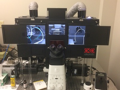
Nikon Eclipse Ti2-E TIRF Microscope
TIRF microscope SOP
Motorized inverted microscope with 25mm field of view and Perfect Focus System 4, motorized epifluorescence turret with following filter cubes: 350, 488, 546, 647 nm and TIRF laser lines 488/561/640 nm.
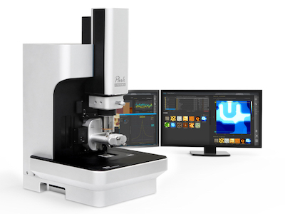
Park Systems NX10 AFM
SOP AFM document
NX10 Atomic Force Microscope (AFM) with Scanning Ion Conductance Microscopy (SICM) module was funded by AFOSR DURIP award FA9550-22-1-0147.
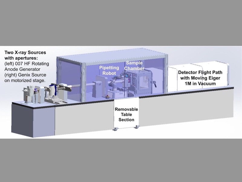
Xenocs Xeuss 3 SAXS/WAXS
More info at MCF website
Automated SAXS/WAXS instrument enables in situ and in operando characterization of structural properties at a variety of length scales. Supported by NSF MRI Award #2117523.
Funders